Overview
 Morton’s Neuroma is the most common neuroma in the foot. It occurs in the forefoot area (the ball of the foot) at the base of the third and fourth toes. It is sometimes referred to as an intermetatarsal neuroma. “Intermetatarsal” describes its location in the ball of the foot between the metatarsal bones (the bones extending from the toes to the midfoot). A neuroma is a thickening, or enlargement, of the nerve as a result of compression or irritation of the nerve. Compression and irritation creates swelling of the nerve, which can eventually lead to permanent nerve damage.
Causes
There are a number of common causes for Morton?s Neuroma, (though the condition can arise spontaneously for reasons still unknown). The Neuroma often occurs in response to irritation, pressure or traumatic injury to one of the digital nerves leading to the toes. A thickening of nerve tissue results as part of the body?s response to the irritation or injury. Abnormal foot movement used to compensate for bunions, hammertoes, flatfeet and other conditions can lead to irritation and development of Morton?s Neuroma. Pronation of the foot may cause the heads of the metatarsal bones to rotate slightly, thereby pinching the nerve running between the metatarsal heads. Chronic pressure or pinching causes the nerve sheath to enlarge, becoming increasingly squeezed, producing worsening pain over time, if not addressed. Morton?s Neuroma can be exacerbated when tight shoes providing little room for the forefoot are worn. Activities which over-pronate the foot (such as walking barefoot in sand) may increase the pain associated with Morton?s Neuroma, as will any high-impact activity, such as jogging.
Symptoms
Morton’s neuroma may cause Burning, pain, tingling, and numbness often shooting into the toes. Discomfort that is worse while walking. Feeling of a lump between the toes. Symptoms are usually temporarily relieved when taking off shoes, flexing toes or rubbing feet.
Diagnosis
To confirm the diagnosis, your doctor will examine your feet. He or she will look for areas of tenderness, swelling, calluses, numbness, muscle weakness and limited motion. To check for a Morton’s neuroma, your doctor will squeeze the sides of your foot. Squeezing should compress the neuroma and trigger your typical pain. In some cases, your doctor will find numbness in the webbed area between the affected toes. Pain in two or more locations on one foot, such as between both the second and third toes and the third and fourth toes, more likely indicates that the toe joints are inflamed rather than a Morton’ neuroma.
Non Surgical Treatment
If you have Morton’s neuroma, shoes with a wider toe area may be recommended. You can also take painkillers to help ease the pain. Steroid injections may also be given to treat the affected nerve. If these treatments don’t work, surgery may be needed. This involves removing the thickened tissue around the nerve (and sometimes the nerve itself) to release the pressure.
Surgical Treatment
When early treatments fail and the neuroma progresses past the threshold for such options, podiatric surgery may become necessary. The procedure, which removes the inflamed and enlarged nerve, can usually be conducted on an outpatient basis, with a recovery time that is often just a few weeks. Your podiatric physician will thoroughly describe the surgical procedures to be used and the results you can expect. Any pain following surgery is easily managed with medications prescribed by your podiatrist.


 Overview
 Symptoms
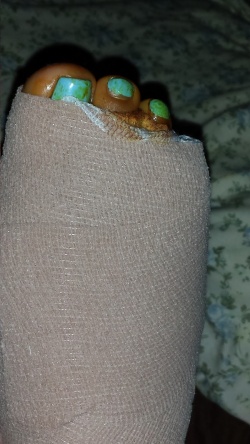 Overview
 Symptoms
 Prevention


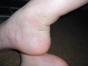 Tendinitis is inflammation of a tendon. Thus, Achilles tendinitis is an inflammation of the Achilles tendon. The inflammation may be localized to the end of the tendon closest to the heel or may spread upward to affect even the muscles of the calf. Swelling often occurs and pain is felt upon contraction of the calf muscles. In severe cases, pain may be felt even at rest. Generally, Achilles tendinitis begins as a dull pain at the back of the lower leg just above the heel when pushing off the ground with the foot. Unless the activity is stopped, the condition rapidly gets worse until any activity requiring a push-off from the ground by the foot becomes quite painful and nearly impossible. If it is left untreated, it can develop into one of two more serious conditions – Achilles tendinosis and Insertional Calcific Tendinitis.
